
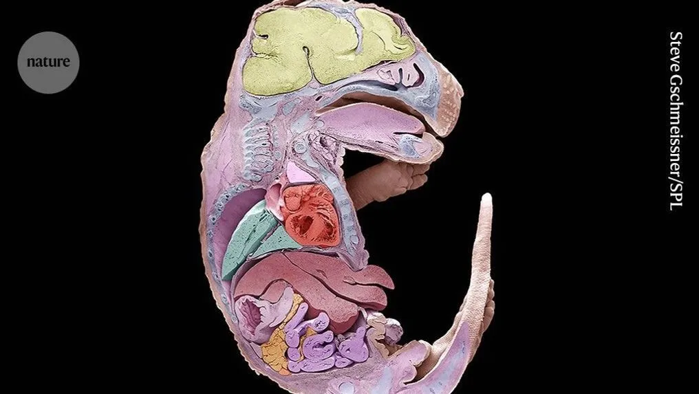
Scientists have achieved a long standing goal in developmental biology by creating a high resolution map of how a mouse grows from a single fertilized egg into a complex organism. Two separate research teams, publishing their work in the journals Science and Cell, utilized cutting edge gene editing to track millions of individual cells as they divided and specialized. This breakthrough allows researchers to see exactly how an embryo organizes itself, providing a window into the chaotic but coordinated process of building a living mammal.
For decades, biologists dreamed of recreating the meticulous mapping done in the 1980s with tiny transparent worms, where every single cell division could be watched under a microscope. However, mammalian development happens behind closed doors and involves billions of cells influenced by unpredictable environmental cues. Jay Shendure of the University of Washington noted that while humans generally share the same basic form, the actual path each cell takes to become part of a lung or a liver varies wildly, even between identical twins.
To solve this puzzle without harming the delicate embryos, the researchers moved beyond standard CRISPR tools toward a more refined technique known as prime editing. Shendure’s team employed a method they call DNA Typewriter, which essentially leaves permanent genetic stamps on cells as they multiply. By reading these indelible marks later on, they were able to reconstruct the ancestral relationships of roughly 1.3 million cells within a two week old embryo.
This ability to record cellular history offers profound insights into how nature maintains consistency despite inherent randomness. By tracing these lineages back to their origin, scientists can better understand why certain tissues form correctly while others may fail during development. It represents a massive leap forward in our understanding of genetics, turning the invisible process of embryonic growth into a readable data set for the first time.